Friday, August 30, 2013

NEW REVOLUTIONS IN MEDICAL SCIENCE

1.Bee Venom Kills HIV:
Nanoparticles carrying a toxin found in bee venom can destroy human immunodeficiency virus (HIV) while leaving surrounding cells unharmed, researchers at Washington University School of Medicine in St. Louis have shown. The finding is an important step toward developing a vaginal gel that may prevent the spread of HIV, the virus that causes AIDS.

Saturday, August 24, 2013

important vedios

1.urea cycle

Sunday, August 18, 2013

lab spotting

1.http://www.slideshare.net/monikasharma7739/family-planningppt

Types of joints movements:
Flexion: Bending parts at a joint so that the angle between them decreases and the parts come closer together (bending the lower limb at the knee).
Extension: Straightening parts at a joint so that the angle between them increases and the parts move farther apart (straightening the lower limb at the knee).
Hyperextension: Excess extension of the parts at a joint, beyond the anatomical position (bending the head back beyond the upright position).
Dorsiflexion: Bending the foot at the ankle toward the shin (bending the foot upward).
Plantar flexion: Bending the foot at the ankle toward the sole (bending the foot downward).
Abduction: Moving a part away from the midline (lifting the upper limb horizontally to form a right angle with he side of the body).
Adduction: Moving a part toward the midline (returning the upper limb from the horizontal position to the side of the body).
Rotation: Moving a part around an axis (twisting the head from side to side). Medial rotation involves movement toward the midline, whereas lateral rotation involves movement in the opposite direction.
Circumduction: Moving a part so that its end follows a circular path (moving the finger in a circular motion without moving the hand).
Supination: Turning the hand so the palm is upward or facing anteriorly (in anatomical position).

Saturday, August 17, 2013

study matter for pharm-d 1st year

1.lipid profile tests
                                                       Lipoprotein structure (chylomicron)
                        ApoA, ApoB, ApoC, ApoE (apolipoproteins); T(triacylglycerol); C (cholesterol); green (phospholipids)
lipoprotein is a biochemical assembly that contains both proteins and lipids, bound to the proteins, which allow fats to move through the water inside and outside cells. The proteins serve to emulsify the lipid (otherwise called fat) molecules. Many enzymes,transporters, structural proteins, antigensadhesins, and toxins are lipoproteins. Examples include the plasma lipoprotein particles classified under high-density(HDL) and low-density (LDL) lipoproteins, which enable fats to be carried in the bloodstream, the transmembrane proteins of the mitochondrion and the chloroplast, and bacterial lipoproteins.
functions
The handling of lipoprotein particles in the body is referred to as lipoprotein particle metabolism. It is divided into two pathways, exogenous and endogenous, depending in large part on whether the lipoprotein particles in question are composed chiefly of dietary (exogenous) lipids or whether they originated in the liver (endogenous), through de novo synthesis of triacylglycerols.
The hepatocytes are the main platform for the handling of TGs and cholesterol; the liver can also store certain amounts of glycogen and triacylglycerols. Intriguingly, adipocytes, though being the main storage cells for triacylglycerols, do not produce any kind of lipoprotein particle.
http://www.srmuniv.ac.in/downloads/Lipid_Profile_Tests.pdf

2.nucleic acid metabolism

3. 4. 5. 6. 7. 8. 9. 10. 11.

12.digestive system ppt. 


13.respiratory system ppt.

14.urinary system ppt.

15.sports and physiology
http://www.dikseo.teimes.gr/spoudastirio/E-NOTES/P/Performance_Enhancing_Drugs_Viewpoints.pdf

16.RADIO IMMUNO ASSAYRadio Immuno m

DEFINITION:a sensitive immunological assay, making use of antibodies and radioactive labelling, for the detection and quantification of biologically important substances, such as hormone concentrations in the blood.

The technique of radioimmunoassay has revolutionized research and clinical practice in many areas, e.g.,
  • blood banking
  • diagnosis of allergies
  • endocrinology
The technique was introduced in 1960 by Berson and Yalow as an assay for the concentration of insulin in plasma. It represented the first time that hormone levels in the blood could be detected by an in vitro assay.

The Technique

  • A mixture is prepared of
    • radioactive antigen
      • Because of the ease with which iodine atoms can be introduced into tyrosine residues in a protein, the radioactive isotopes 125I or 131I are often used.
    • antibodies ("First" antibody) against that antigen.
  • Known amounts of unlabeled ("cold") antigen are added to samples of the mixture. These compete for the binding sites of the antibodies.
  • At increasing concentrations of unlabeled antigen, an increasing amount of radioactive antigen is displaced from the antibody molecules.
  • The antibody-bound antigen is separated (see below) from the free antigen in the supernatant fluid, and
  • the radioactivity of each is measured.
  • From these data, a standard binding curve, like this one shown in red, can be drawn.
  • The samples to be assayed (the unknowns) are run in parallel.
  • After determining the ratio of bound to free antigen ("cpm Bound/cpm Free") in each unknown, the antigen concentrations can be read directly from the standard curve (as shown above).
  • Separating Bound from Free Antigen

    There are several ways of doing this.
    • Precipitate the antigen-antibody complexes by adding a "second" antibody directed against the first. For example, if a rabbit IgG is used to bind the antigen, the complex can be precipitated by adding an antirabbit-IgG antiserum (e.g., raised by immunizing a goat with rabbit IgG). This is the method shown in the diagram above.
    • The antigen-specific antibodies can be coupled to the inner walls of a test tube [View another example]. After incubation,
      • the contents ("free") are removed;
      • the tube is washed ("bound"), and
      • the radioactive of both is measured.
    • The antigen-specific antibodies can be coupled to particles, like Sephadex. Centrifugation of the reaction mixture separates
      • the bound counts (in the pellet) from
      • the free counts in the supernatant fluid.
    Radioimmunoassay is widely-used because of its great sensitivity. Using antibodies of high affinity (K0 = 108–1011 M−1), it is possible to detect a few picograms (10−12 g) of antigen in the tube.
  • The greater the specificity of the antiserum, the greater the specificity of the assay.
  • The main drawbacks to radioimmunoassay are the expense and hazards of preparing and handling the radioactive antigen.
    • Both 125I or 131I emit gamma radiation that requires special counting equipment;
    • The body concentrates iodine atoms — radioactive or not — in the thyroid gland where they are incorporated in thyroxine (T4).

    • Despite these drawbacks, RIA has become a major tool in the clinical laboratory where it is used to assay
      • plasma levels of:
        • most of our hormones;
        • digitoxin or digoxin in patients receiving these drugs;
        • certain abused drugs
for the presence of hepatitis B surface antigen (HBsAg) in donated blood;anti-DNA antibodies in systemic lupus erythematosus (SLE).

17.ELISA(enzyme-linked immunosorbent assay)
Definition:A sensitive immunoassay that uses an enzyme linked to an antibody or antigen as a marker for the detection of a specific protein, especially an antigen or antibody. It is often used as a diagnostic test to determine exposure to a particular infectious agent, such as the AIDS virus, by identifying antibodies present in a blood sample.
                                                        A 96-well microtiter plate used in elisa
ELISAs are performed in 96-well plates which permits high throughput results. The bottom of each well is coated with a protein to which will bind the antibody you want to measure. Whole blood is allowed to clot and the cells are centrifuged out to obtain the clear serum with antibodies (called primary antibodies). The serum is incubated in a well, and each well contains a different serum (see figure below). A positive control serum and a negative control serum would be included among the 96 samples being tested.

Main ELISA types

*Direct ELISA

- direct detection (primary antibody is conjugated with the enzyme/tag) 
*Indirect ELISA – signal amplification
- indirect detection (labelling the secondary antibody - produced
against the primary antibody type; e.g. anti-mouse IgG, a rabbit Ab)
*Sandwich ELISA – more efficient signal amplification
- indirect antigen immobilization (by capture antibody)
- indirect detection (labelled secondary antibody)
*Competitive ELISA
A third use of ELISA is through competitive binding. The steps for this ELISA are somewhat different from the first two examples:
  1. Unlabeled antibody is incubated in the presence of its antigen (sample).
  2. These bound antibody/antigen complexes are then added to an antigen-coated well.
  3. The plate is washed, so unbound antibody is removed. (The more antigen in the sample, the less antibody will be able to bind to the antigen in the well, hence "competition".)
  4. The secondary antibody, specific to the primary antibody, is added. This second antibody is coupled to the enzyme.
  5. A substrate is added, and remaining enzymes elicit a chromogenic or fluorescent signal.
  6. The reaction is stopped to prevent eventual saturation of the signal.
A "sandwich" ELISA, is used to detect sample antigen. The steps are:
  1. A surface is prepared to which a known quantity of capture antibody is bound.
  2. Any nonspecific binding sites on the surface are blocked.
  3. The antigen-containing sample is applied to the plate.
  4. The plate is washed to remove unbound antigen.
  5. A specific antibody is added, and binds to antigen (hence the 'sandwich': the Ag is stuck between two antibodies)
  6. Enzyme-linked secondary antibodies are applied as detection antibodies that also bind specifically to the antibody's Fc region (nonspecific).
  7. The plate is washed to remove the unbound antibody-enzyme conjugates.
  8. A chemical is added to be converted by the enzyme into a color or fluorescent or electrochemical signal.
  9. The absorbency or fluorescence or electrochemical signal (e.g., current) of the plate wells is measured to determine the presence and quantity of antigen.
For additional info please check the link:http://www3.szote.u-szeged.hu/hurodocs/downloads/biochemistry/lectures/ELISA_lecture_HURO_Keresztes_final_small%20.pdf

18.Active Transport vs Passive Transport
Active and passive transport are biological processes that move oxygen, water and nutrients into cells and remove waste products. Active transport requires chemical energy because it is the movement of biochemicals from areas of lower concentration to areas of higher concentration. On the other hand, passive trasport moves biochemicals from areas of high concentration to areas of low concentration; so it does not require energy.

Wednesday, August 14, 2013

INTERESTING FACTS


1.Foot Reflexology 
Massage the part of your foot that corresponds to the part of your body that hurts and it will slowly reduce and eventually get rid of pain. Helps promote blood circulation to that particular part of the body.

                                                                                  
2.12 signs of breast cancer revealed


3.bp normal level

4.site for insulin injection

5.HEALTH IMPORTANT TIPS

6.ARE YOU RIGHT OR LEFT BRAINED?
7.symbols in pharmacy
8.FACTS OF BLOOD TYPE


Saturday, August 10, 2013

INJECTIONS


Intradermal Injections

This technique involves the injection of the fluids into the top layer of the skin, which is soft and pliable. Mainly used for treating certain health problems , including many allergies and tuberculosis. The liquid medicine is inserted with an intradermal injection, which will lie just beneath the skin surface in between the layers of skin. The needle is extremely tiny, and it inserts the fluid properly under the surface of the skin. 

Intramuscular Injections

This is the most common way of injecting medication directly into a patient. For rapid absorption of the medicine this is a very useful process because the medicine from this injection is inserted directly into the muscle. This allows the medicine to gain easy access to the blood stream and quickly begin its healing work. Intramuscular injections are the best and the safest way of injecting medication into a patient.

Subcutaneous Injections

Such type of injections are used where the medicine needs to be absorbed slowly. In this type of injection, the needle has to go through the first 2 layers of skin that is the epidermis and dermis. The needle should further penetrate into the fatty layer of the skin, known as the subcutaneous tissue. Medicines administered through subcutaneous injections have the least chances of having an adverse reaction. Insulin is one type of medicine that is injected in this way, so also a number of immunizations. 
In all these injections, the size of the needle varies. The deeper the injection, the longer the needle should be. In intramuscular injection, the needle is at least a few inches long. Subcutaneous injections have needles which are approximately an inch long. Intradermal injections use the shortest needle because they are only inserted just beneath the first layer of skin. 

List of Injections


Dosage form


Banned lethal drugs freely available


Thursday, August 8, 2013

PHARM-D

Doctor of Pharmacy (Pharm.D.) is a professional doctor degree in pharmacy. In some countries, it is a first professional degree, and a prerequisite for licensing to exercise the profession of PharmacistPharm.D course introduced by the Government of India and the Pharmacy Council of India in 2008. The Pharm.D program is a pre-PhD, post-graduate professional doctoral program. It was introduced to improve clinical pharmacy services in India and it is the only pharmacy service which is in direct contact with patient health care system. The first batch of Pharm.D post baccalaureate students graduated in August 2011. The Pharm.D degree requires five years of classroom and hospital based didactic study (two years didactic post-baccalaureate course), followed by one year of internship training in addition to ongoing practicals and research project. The entry requirements for this 6 year pharmD program is after 12 years of school education with maths & science sub: or diploma in pharmacy and for post baccalaureate entry is after 4 year B.Pharm undergraduate qualification. In india pharm D students and graduates are actively participating in direct patient care in hospitals. Pharm D graduation is a post graduate professional doctoral degree in india. With reference to Clarification on Pharm.D qualification, it is clarified to all universities that Pharm.D is a post graduate degree and passed out students can directly register for Ph.D From 2012. PharmD is a registered teaching degree for D.pharm, B.pharm, M.pharm & PharmD courses. the Amendment passed by PCI (Pharmacists' professional association called Pharmacy Council of India) states that the person can use the initials "Dr" before their name after completion of their pharm-D.


Drug incompatability

course syllabus


Tuesday, August 6, 2013

BASICS

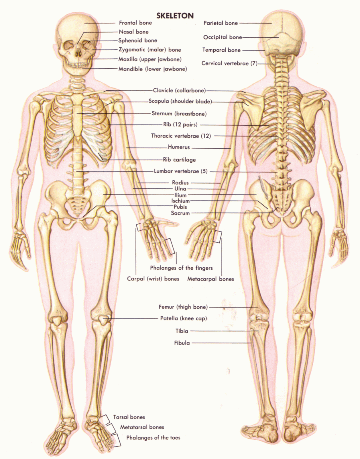
BONES IN HUMAN BODY

Fun Facts About Bones

  • The adult human body has 206 of them. 
  • There are 26 bones in the human foot. 
  • The human hand, including the wrist, contains 54 bones. 
  • The femur, or thighbone, is the longest and strongest bone of the human skeleton. 
  • The stapes, in the middle ear, is the smallest and lightest bone of the human skeleton. 
  • Arms are among the most commonly broken bones, accounting for almost half of all adults' broken bones. The collarbone is the most commonly broken bone among children. 
  • Bones stop growing in length during puberty. Bone density and strength will change over the course of life, however. 
  • The only bone in the human body not connected to another is the hyoid, a V-shaped bone located at the base of the tongue. 
  • Bones are made up of calcium, phosphorus, sodium, and other minerals, as well as the protein collagen. 
  • Bones function as the skeleton of the human body, allow body parts to move and protect organs from impact damage. They also produce red and white blood cells.

  •                          

                                 Fun Facts About Joints


    • Joints are the place where two bones meet or connectJoints are the place where two bones meet or connect. 
    • Ligaments are short bands of tough fibrous connective tissue that function to connect one bone to another, forming the joint. 
    • Tendons are made of elastic tissue and also play a key role in the functioning of joints. They connect muscle to bone. 
    • A coating of another fibrous tissue called cartilage covers the bone surface and keeps the bones from rubbing directly against each other. 
    • Some joints move and some don't. Joints in the skull don't move. Synovial joints are movable joints. They make up most of the joints in the body and are located mostly in the limbs, where mobility is critical. They contain synovial fluid, which helps them to move freely. 
    • Ball and socket joints, such as hip and shoulder joints, are the most mobile type of joint. They allow you to move your arms and legs in many different directions. 
    • Ellipsoidal joints, such as the one at the base of the index finger, allow bending and extending. 
    • Gliding joints are found between flat bones that are held together by ligaments. Some bones in the wrists and ankles move by gliding against each other. 
    • Hinge joints are those in the knee and elbow. They enable movement similar to the way a hinged door moves. 
    • The pivot joint in the neck allows it to turn from side to side.